Osteosarcoma
Epidemiology
- Osteosarcomas usually involve the long bones and are most commonly diagnosed during childhood.
- However, head and neck osteosarcomas are mostly found in adults and these tumors account for 7 to 1 6% of all osteosarcomas.
- The most common site in the head and neck is the mandible.
- Skull base tumors are rare
- . There is a known relationship between the development of osteosarcoma with Paget’s disease, fibrous dysplasia, bilateral retinoblastoma, and irradiation.
Clinical Findings
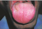
- Patients with osteosarcoma usually present with a mass with or without pain.
- Most of these tumors are large at presentation. Clinically, the mass is rock hard.
- Lesions located in the skull base or deep face may have less obvious mass effect or facial deformity.
Pathology
- The cell of origin is the osteoblast. Osteosarcomas are very pleomorphic. If they are well differentiated, osteoid formation is evident.
- Osteoids may show variable calcification or ossification.
- When these lesions breach the bone cortex, they lift the periosteum.
- New bone with the typical sunray appearance may form beneath the periosteum.
- The tumor frequently metastasizes and the most common site for secondaries is the lung.
Treatment
- Treatment usually involves a combination of surgery, radiation therapy, and chemotherapy.
- The tumor shows very high frequencies of local recurrence if only surgery is performed.
Imaging Findings
CT
- The CT appearances are variable depending on the degree of calcification and ossification of tumor osteoid.
- CT may show a tumor comprising mainly dense bone or a large soft tissue mass with associated bone destruction.
- The presence of sunray spiculation is characteristic
MR
- Like CT, the MR appearances depend on the degree of calcification and ossification.
- Densely mineralized lesions appear as signal void areas.
- The soft tissue component enhances well and T2-weighted images show high signals.
- Associated marrow infiltration is best delineated with T 1 -weighted images where high signal intensity marrow is replaced by intermediate signal intensity neoplastic tissue
Imaging Pearls
- When an osteosarcoma is suspected, it may be useful to find out if there is a history of previous radiation therapy.
- There are several criteria for the diagnosis of radiation-associated sarcoma:
(a) there is a history of radiation therapy
(b) the second neoplasm must occur within the field of radiation
(c) the histology of the second neoplasm must be distinctly different from that of the primary tumor
(d) a latency period of many years, arbitrarily taken to be at least 5 years, must have lapsed between radiation therapy and the occurrence of the second tumor


ــــــــــــــــــــ► ⒹⒺⓃⓉⒶⓁ–ⓈⒸⒾⒺⓝⓒⒺ ◄ــــــــــــــــــــ


seize your rss feed as I can not to find your email subscription link or newsletter service. Do you’ve any?
you can subscribe now from the box on the right side of our website