Rhabdomyosarcoma
Epidemiology
- Rhabdomyosarcomas are most commonly seen in the pediatric age group. This malignant neoplasm is also the most common soft tissue tumor in childhood.
- It accounts for about 50 to 70% of all childhood sarcomas.
- Approximately 30 to 40% occur in the head and neck.
- About half of patients present before the age of 5 years.
- There is no sex predilection.
Clinical Findings
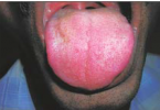
- Signs and symptoms depend on the size and location of the lesion.
- Large tumors often cause trismus as a result of masticator muscle dysfunction or involvement of the temporomandibular joint.
- Pain may be related to bone erosion or nerve involvement.
- Physical examination reveals medial displacement of the pharyngeal mucosa.
- Patients may also present with nasal stuffiness if the tumor extends into the maxillary sinus or nasal cavity.
- These complaints may be similar to those of a juvenile angiofibroma.
Pathology
- There are four histologic subtypes. The well-differentiated pleomorphic type is most commonly seen in adults.
- The alveolar type predominates in the adolescent age group.
- The embryonal and botryoid types are typically encountered in childhood.
- Besides the masticator space, other common sites include the orbit, nasopharynx, mastoid and middle ear, and the sino nasal area.
- The tumor spreads by both lymphatic and hematogenous routes.
- Metastasis to the lungs, bones, and marrow is common.
- In 8% of patients regional nodal spread can be seen.
Treatment
- Rhabdomyosarcoma is best treated using a multimodality approach including surgery, radiation therapy, and chemotherapy.
- The best prognosis is seen in orbital rhabdomyosarcomas.
- Tumors that involve the nasopharynx and sinonasal regions have a propensity to extend intracranially and tend to have the worst prognosis.
- However, the prognosis has improved over the years with an overall 3-year survival of 85% and a 5-year disease free rate approaching 73%.
Imaging Findings
CT
- The tumor is usually large at presentation.
- Contrast-enhanced CT usually shows mild enhancement.
- These tumors may extend anteriorly into the maxillary sinus and demonstrate aggressive destruction of the posterior wall of the sinus.
- Advanced lesions may also demonstrate aggressive bone destruction of the mandible and skull base.
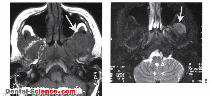
MR
- These locally aggressive tumors are an intermediate signal on T l -weighted sequences and ahigh signal on T2W .
- The tumors homogeneously enhance following intravenous contrast.
- The internal characteristics tend to be homogeneous without internal flow voids.
Imaging Pearls
• A rhabdomyosarcoma should be the initial consideration for a primary rumor arising within the masticator space in a child .
• The lack of flow voids and the presence of aggressive bone desrruction help differentiate a rhabdomyosarcoma from a juvenile angiofibroma in an adolescent male that extends into the sinonasal region.

ــــــــــــــــــــ► ⒹⒺⓃⓉⒶⓁ–ⓈⒸⒾⒺⓝⓒⒺ ◄ــــــــــــــــــــ