PTERYGOPALATINE FOSSA
1)Overview and Topographic Anatomy
GENERAL INFORMATION
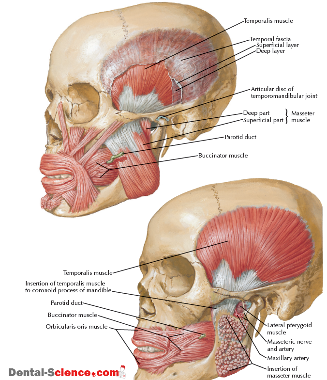
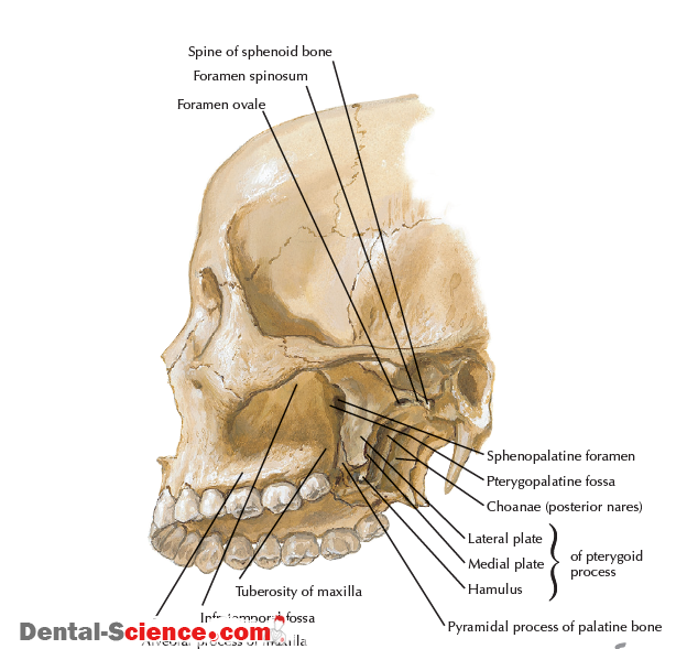
Pyramid-shaped fossa on the lateral aspect of the skull between the maxilla’s infratemporal surface and the pterygoid process of the sphenoid Contains major nerves and blood vessels that supply the nasal cavity, upper jaw, hard palate, and soft palate: the maxillary division of the trigeminal nerve, pterygopalatine (sphenopalatine, Meckel’s) ganglion, and 3rd portion of the maxillary artery Allows the infratemporal fossa, middle cranial fossa, foramen lacerum, nasopharynx, nasal cavity, orbital cavity, and oral cavity to communicate 7 foramina/fissures allow passage of nerves and vessels
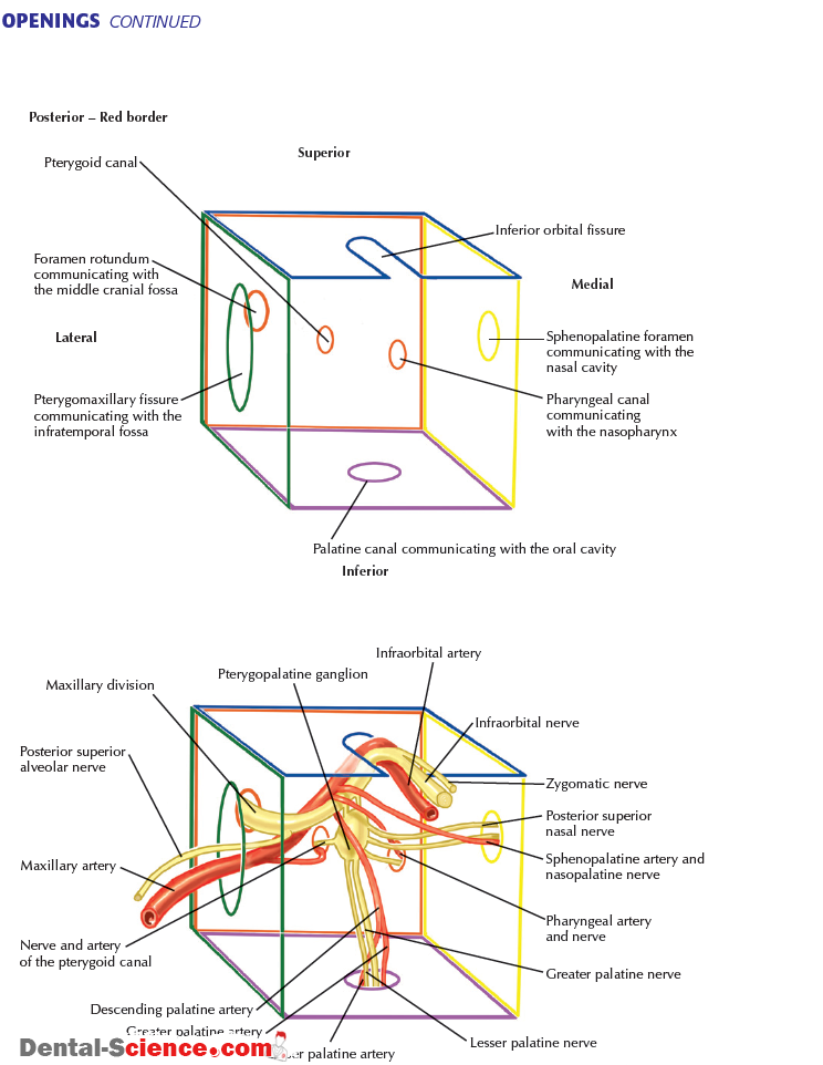
2)Borders and Opening :
Border | Structures |
Anterior wall | Infratemporal surface of the maxilla |
Posterior wall | Pterygoid process of the sphenoid |
Medial wall | Perpendicular plate of the palatine |
Lateral wall | None (open to the pterygomaxillary fissure) |
Superior wall | Inferior surface of the sphenoid and the orbital plate of the palatine bone |
Inferior wall | Pyramidal process of the palatine |
Opening | Location | Transmitted Structures |
Pterygomaxillary fissure | Lateral part of the pterygopalatine fossa Between the infratemporal fossa and the pterygopalatine fossa | Posterior superior alveolar n. from the pterygopalatine fossa into the infratemporal fossa 3rd part of the maxillary a. from the infratemporal fossa into the pterygopalatine fossa A variable network of veins, such as the sphenopalatine, into the pterygoid plexus of vv. |
Sphenopalatine foramen | Medial wall of the pterygopalatine fossa Between the nasal cavity and the pterygopalatine fossa Often located posterior to the middle nasal concha | Nasopalatine n. Posterior superior nasal nn. Sphenopalatine vessels |
Inferior orbital fissure | Superior part of the pterygopalatine fossa Between the pterygopalatine fossa and the orbit Continues posteriorly with the superior part of the pterygomaxillary fissure | Infraorbital n. from the maxillary division of the trigeminal n. Zygomatic n. from the maxillary division of the trigeminal Infraorbital vessels Inferior ophthalmic v. that connects with the pterygoid plexus of veins |
Palatine canal | Inferior part of the pterygopalatine fossa Between the pterygopalatine fossa and the hard and the soft palate Eventually terminates into the greater and lesser palatine foramina | Greater palatine n. and vessels (through the greater palatine foramen) onto the hard palate Lesser palatine n. and vessels (through the lesser palatine foramen) onto the soft palate |
Foramen rotundum | Posterolateral part of the pterygopalatine fossa Between the pterygopalatine fossa and the middle cranial fossa | Maxillary division of the trigeminal n. |
Pterygoid canal | Posterior part of the pterygopalatine fossa Between the pterygopalatine fossa and the foramen lacerum Inferior and medial to the foramen rotundum | Nerve of the pterygoid canal (vidian n.) An accompanying artery |
Pharyngeal canal | Posteromedial part of the pterygopalatine fossa Between the pterygopalatine fossa and the nasopharynx Medial to the pterygoid canal | Pharyngeal n. Pharyngeal vessels |

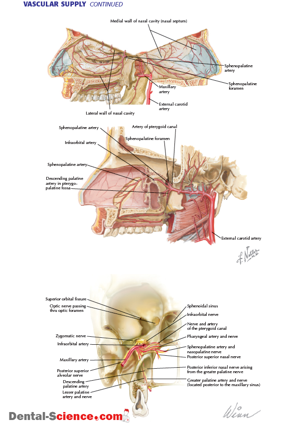
3)Arterial Supply :
ARTERIAL SUPPLY | |||
Artery | Source | Course | |
Maxillary (3rd part) | External carotid a. | Passes from the infratemporal fossa into the pterygopalatine fossa via the pterygomaxillary fissure Prior to passing through the pterygomaxillary fissure, it gives off the posterior superior alveolar a. (the only artery from the 3rd part of the maxillary a. that does not normally branch off within the pterygopalatine fossa) | |
Infraorbital | The continuation of the 3rd part of the maxillary a. | Accompanied by the infraorbital n. and v. The artery passes forward in the infraorbital groove, infraorbital canal, and exits the infraorbital foramen In the infraorbital canal, it gives rise to various orbital branches that aid in supplying the lacrimal gland and extraocular muscles In the infraorbital canal, it also gives rise to the anterior and middle (if present) superior alveolar aa. that supply the maxillary teeth from the central incisors to the premolars (where they anastomose with the posterior superior alveolar a.) and the mucous membrane of the maxillary sinus On exiting the infraorbital foramen, the artery is located between the levator labii superioris and levator anguli oris mm. and follows the branching pattern of the nerve: ● Inferior palpebral branch (supplies the lower eyelid) ● Nasal branch (supplies the lateral side of the nose) ● Superior labial branch (supplies the upper lip) | |
Descending palatine | 3rd part of the maxillary a. | Descends into the palatine canal Within the canal, the artery splits into the greater and lesser palatine aa. Greater palatine a. exits the greater palatine foramen and passes anteriorly toward the incisive foramen and supplies the hard palate gingiva, mucosa, and palatal glands and anastomoses with the terminal branch of the sphenopalatine a. that exits the incisive foramen Lesser palatine a. supplies the soft palate and palatine tonsil | |
Artery of the pterygoid canal | Passes posteriorly into the pterygoid canal, accompanying the nerve of the pterygoid canal (vidian n.) Helps supply the auditory tube and sphenoid sinus | ||
Pharyngeal | Passes posteromedially into the pharyngeal canal Helps supply the auditory tube and nasopharynx | ||
Sphenopalatine | Passes medially into the sphenopalatine foramen to enter the nasal cavity It then gives rise to the posterior lateral nasal branches and posterior septal branches, which supply the nasal concha, mucous membranes, and nasal septum The sphenopalatine a. continues along the nasal septum to enter the hard palate via the incisive canal | ||
4)Venous Supply :
VENOUS DRAINAGE | ||
Vein | Course | |
Posterior superior alveolar | Receives blood from the posterior teeth and soft tissue | Eventually communicate with the pterygoid plexus of veins |
Pharyngeal | Receives blood from the nasopharynx | |
Descending palatine | Receives blood from the hard and soft palate | |
Infraorbital | Receives blood from the midface via the lower eyelid, lateral side of the nose, and the upper lip | |
Sphenopalatine | Receives blood from the nasal cavity and the nasal septum | |
Vein of the pterygoid canal | Receives blood from the foramen lacerum region and the sphenoid sinus | |
Inferior ophthalmic | Receives blood from the floor of the orbit Branches into 2 parts The first branch travels posteriorly with the infraorbital v. that passes through the inferior orbital fissure to communicate with the pterygoid plexus and the cavernous sinus The main branch travels posteriorly to communicate with the superior ophthalmic vein in the superior orbital fissure or travels posteriorly in the fissure to join the cavernous sinus | |
Pterygoid plexus | An extensive network of veins that parallels the 2nd and 3rd parts of the maxillary a. The tributaries of the pterygoid plexus eventually converge to form a short maxillary v. | |

5)Nerve Supply :
MAXILLARY NERVE | ||
Nerve | Source | Course |
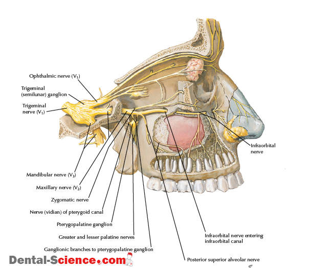
Maxillary division of the trigeminal n. | Trigeminal n. | Sensory in function Travels along the lateral wall of the cavernous sinus Before exiting the middle cranial fossa, it gives off a meningeal branch that innervates the dura mater Passes from the middle cranial fossa into the pterygopalatine fossa via the foramen rotundum Within the pterygopalatine fossa, gives rise to 4 branches: ● Posterior superior alveolar n. ● Zygomatic n. ● Ganglionic branches ● Infraorbital n. |
Posterior superior alveolar | Maxillary division of the trigeminal n. in pterygopalatine fossa | Passes through the pterygomaxillary fissure to enter the infratemporal fossa In the infratemporal fossa, it passes on the posterior surface of the maxilla along the region of the maxillary tuberosity Gives rise to a gingival branch that innervates the buccal gingiva alongside the maxillary molars Enters the posterior surface of the maxilla and supplies the maxillary sinus and the maxillary molars with the possible exception of the mesiobuccal root of the 1st maxillary molar |
Zygomatic | Passes through the inferior orbital fissure to enter the orbit Passes on the lateral wall of the orbit and branches into the zygomaticotemporal and zygomaticofacial branches A communicating branch from it joins the lacrimal n. from the ophthalmic division of the trigeminal to carry autonomics to the lacrimal gland | |
Ganglionic branches |
| Usually 1 or 2 ganglionic branches that connect the maxillary division of the trigeminal to the pterygopalatine ganglion Contain sensory fibers that pass through the pterygopalatine ganglion (without synapsing) to be distributed with the nerves that arise from the pterygopalatine ganglion Also contain postganglionic autonomic fibers to the lacrimal gland that pass through the pterygopalatine ganglion (Parasympathetic fibers form a synapse here between the preganglionic fibers from the vidian n. and the postganglionic fibers)
|
Infraorbital | Considered the continuation of the maxillary division of the trigeminal n. | Passes through the inferior orbital fissure to enter the orbit Passes anteriorly through the infraorbital groove, infraorbital canal, and exits onto the face via the infraorbital foramen Within the infraorbital canal, it gives rise to: ● Anterior superior alveolar (supplies the maxillary sinus; maxillary central incisor, lateral incisor, and canine; gingiva and mucosa alongside the same teeth) ● A small branch of the anterior superior alveolar (supplies the nasal cavity) ● Middle superior alveolar (present about 70% of the time; supplies the maxillary sinus, maxillary premolars and often the mesiobuccal root of the 1st maxillary molar, and gingiva and mucosa alongside the same teeth) |
BRANCHES OF THE MAXILLARY DIVISION OF THE TRIGEMINAL NERVE | ||
ASSOCIATED WITH THE PTERYGOPALATINE GANGLION | ||
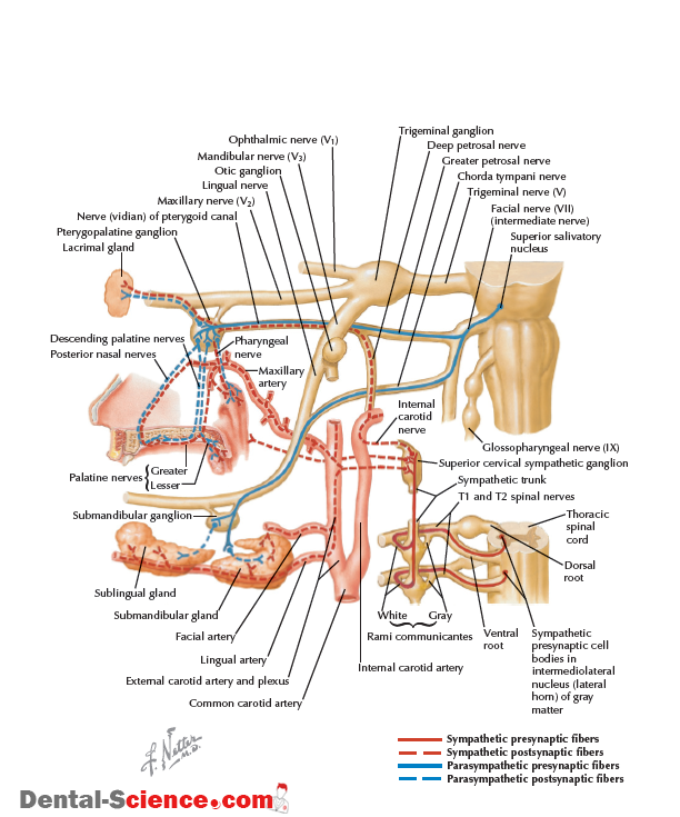
A parasympathetic ganglion named because it is a collection of cell bodies in the peripheral nervous system (postganglionic cell bodies) The ganglionic branches are of the maxillary division of the trigeminal n. that pass through the pterygopalatine ganglion The vidian n. connects to the pterygopalatine ganglion 3 sets of nerve fibers travel through the pterygopalatine ganglion: ● General sensory fibers from the trigeminal n. (without synapsing) ● Postganglionic sympathetic fibers (carried to the pterygopalatine ganglion via the vidian n, without synapsing) ● Preganglionic parasympathetic fibers (carried to the pterygopalatine ganglion via the vidian n. and formed by synapsing in the pterygopalatine ganglion with the postganglionic parasympathetic fibers) All branches arising from the pterygopalatine ganglion carry these 3 sets of fibers to the areas where they terminate These nerves of the maxillary division travel through the pterygopalatine ganglion: ● Nasopalatine n. ● Posterior superior nasal n. ● Greater palatine n. ● Lesser palatine n. ● Pharyngeal n. | ||
Branch | Source | Course |
Vidian (nerve of the pterygoid canal) | Formed by the greater and deep petrosal nn. | An autonomic nerve: ● Greater petrosal n. carries the preganglionic parasympathetic fibers ● Deep petrosal n. carries the postganglionic sympathetic fibers Communicates with the pterygopalatine ganglion, which allows the autonomics to be distributed along any nerve connected to the ganglion |
Nasopalatine | Branches of the pterygopalatine ganglion in the pterygopalatine fossa | Passes through the sphenopalatine foramen to enter the nasal cavity Passes along the superior portion of the nasal cavity to the nasal septum; then travels anteroinferiorly to the incisive canal Exits the incisive foramen on the hard palate and supplies the palatal gingiva and mucosa from the region of the central incisors to the canines |
Posterior superior nasal | Passes through the sphenopalatine foramen to enter the nasal cavity, where it divides into 2 nerves: ● Lateral posterior superior (supplies the lateral wall of the nasal cavity) ● Medial posterior superior nasal (supplies the posterosuperior portion of the nasal septum) | |
Greater palatine | Passes through the palatine canal to enter the hard palate via the greater palatine foramen Supplies the palatal gingiva and mucosa from the area in the premolar region to the posterior border of the hard palate to the midline | |
Lesser palatine | Passes through the palatine canal to enter and supply the soft palate via the lesser palatine foramen | |
Pharyngeal | Passes through the pharyngeal canal to enter and supply the nasopharynx | |

AUTONOMICS TRAVERSING THE PTERYGOPALATINE FOSSA | |||
Type of Neuron | Name of Cell Body | Characteristics of the Cell Body |
Course of the Neuron |
Anatomic Pathway for Parasympathetics Associated with the Maxillary Division of the Trigeminal Nerve | |||
preganglionic neuron |
Superior salivatory nucleus |
A collection of nerve cell bodies located in the pons Travel through the nervus intermedius of the facial n. into the internal acoustic meatus In the facial canal, the facial n. gives rise to 2 parasympathetic branches: ● Greater petrosal n. ● Chorda tympani n. | Greater Petrosal Nerve
Greater petrosal n. exits along the hiatus for the greater petrosal n. toward the foramen lacerum, where it joins the deep petrosal n. (sympathetics) to form the nerve of the pterygoid canal (vidian n.) Vidian n. passes through the pterygoid canal and enters the pterygopalatine fossa, where it joins with the pterygopalatine ganglion |
Postganglionic neuron | Pterygopalatine ganglion | Pterygopalatine ganglion is a collection of nerve cell bodies located in the pterygopalatine fossa Postganglionic parasympathetic fibers that arise in the pterygopalatine ganglion are distributed to the ophthalmic and maxillary divisions of the trigeminal n. to the: ● Lacrimal gland ● Nasal glands ● Palatine glands ● Pharyngeal glands | Ophthalmic Division Distribution
Postganglionic fibers travel along the zygomatic branch of the maxillary division for a short distance to enter the orbit A short communicating branch joins the lacrimal n. of the ophthalmic division of the trigeminal n. These fibers innervate the lacrimal gland to cause the secretion of tears
Maxillary Division Distribution
Postganglionic fibers travel along the maxillary division of the trigeminal n. to be distributed along its branches that are located in the nasal cavity, oral cavity, and pharynx (e.g., nasopalatine, greater palatine) These fibers innervate: ● Nasal glands ● Palatine glands ● Pharyngeal glands |
AUTONOMICS TRAVERSING THE PTERYGOPALATINE FOSSA CONTINUED | |||||||
Type of Neuron | Name of Cell Body | Characteristics of the Cell Body |
Course of the Neuron | ||||
Anatomic Pathway for Sympathetics Associated with the Maxillary Division of the Trigeminal Nerve | |||||||
Preganglionic neuron |
Intermediolateral horn nucleus |
Collection of nerve cell bodies located in the lateral horn nucleus of the spinal cord between spinal segments T1 and T3 (and possibly T4) |
Arise from the intermediolateral horn nuclei from T1 to T3 (4) Travel through the ventral root of the spinal cord to the spinal n. Enter the sympathetic chain via a white ramus communicantes Once in the sympathetic chain, the preganglionic fibers for the eye will ascend and synapse with postganglionic fibers in the superior cervical ganglion | ||||
Postganglionic neuron | Superior cervical ganglion | Collection of nerve cell bodies located in the superior cervical ganglion, which is located at the base of the skull Postganglionic sympathetic fibers follow the internal carotid or external carotid a. to pass near their respective effector organs (e.g., nasal cavity) | Nasal Cavity and Palate
Postganglionic sympathetic fibers follow both the internal and external carotid aa. Postganglionic sympathetic fibers from the internal carotid branch in the region of the foramen lacerum to form the deep petrosal n. The deep petrosal n. joins the greater petrosal n. (parasympathetics) to form the nerve of the pterygoid canal (vidian n.) Postganglionic sympathetic fibers travel along the branches of the maxillary division of the trigeminal n. associated with the pterygopalatine ganglion to be distributed along its branches in the nasal cavity and palate Postganglionic sympathetic fibers from the external carotid branch and follow the maxillary a. These fibers travel along the branches of the maxillary a. to be distributed along the nasal cavity and palate
Lacrimal Gland
Postganglionic sympathetic fibers follow the internal carotid a. Postganglionic sympathetic fibers from the internal carotid branch off in the region of the foramen lacerum to form the deep petrosal n. | ||||
Postganglionic neuron |
|
|
The deep petrosal n. joins the greater petrosal n. (parasympathetics) to form the nerve of the pterygoid canal (vidian n.) Postganglionic fibers travel along the zygomatic branch of the maxillary division for a short distance to enter the orbit A short communicating branch joins the lacrimal n. of the ophthalmic division of the trigeminal n. These fibers are distributed to the lacrimal gland | ||||

6)Imaging :