Benign Minor Salivary Gland Tumors
Epidemiology
- A minor salivary gland consists of cellular constituents of the major salivary glands (parotid, submandibular, sublingual) that are located within the mucosa of the upper aero digestive tract.
- It has been estimated that there are between 500 and 1 000 minor salivary glands located throughout the oral cavity and oropharynx.
- They may be found within the hard and soft palate, uvula, lips, retromolar trigone, tongue base, floor of mouth, and tonsil.
- The same malignancies that arise in the major salivary glands occur in the minor salivary glands.
- Depending on the series, approximately one half of minor salivary gland tumors are benign as compared with approximately 70 to 80% of parotid tumors that are benign.
Clinical Findings
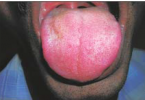
- Patients often present with asymptomatic masses that have been present for several months.
- Pain and ulceration may be present; however, these are not consistent findings.
Pathology
- The benign tumors that constitute minor salivary gland tumors include pleomorphic adenoma, monomorphic adenomas, and Warthin’s tumor.
Treatment
- The exact treatment depends on the pathology.
- For most benign tumors complete local resection is adequate.
Imaging Findings
CT
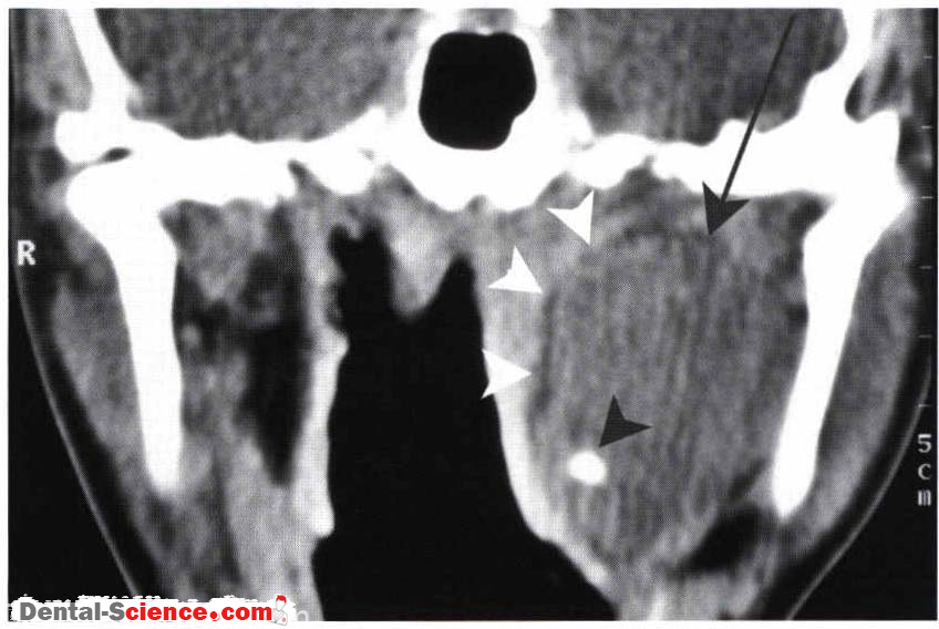
- The CT findings are not specific.
- The presence of regressive remodeling of [he surrounding bone for lesions that arise in the hard palate is suggestive of a benign minor salivary gland tumor
MR
- In general, the imaging findings are nonspecific.
- However, oral cavity or oropharyngeal lesions that are low to intermediate signal on T I -weighted and increased signal on T2- weighted sequences is suggestive of pleomorphic adenoma
Imaging Pearls
- In general, these are uncommon lesions with diagnosis usually at histology following initial biopsy.
- The intent of imaging is to provide information that cannot be detected on clinical examination.
- CT is helpful to evaluate the extent of bone erosion.
- MR should be performed to determine the presence of submucosal spread and deep invasion.

ــــــــــــــــــــ► ⒹⒺⓃⓉⒶⓁ–ⓈⒸⒾⒺⓝⓒⒺ ◄ــــــــــــــــــــ